Understanding a Cyst Foot Bottom: Causes, Diagnosis, and Proven Treatment Options

Discovering an unfamiliar lump on your body can immediately trigger a wave of concern, especially when it appears in a highly functional, weight-bearing area. When patients first notice a bump beneath their soles, they often search for information about a cyst foot bottom to understand whether it is a minor nuisance or a sign of a deeper structural problem. The reality is far less alarming than initial fears suggest. The vast majority of these plantar lumps are benign, fluid-filled sacs known medically as ganglion cysts. While they are far more common on the wrists and hands, their occurrence on the ankle and plantar foot surface is well-documented in orthopedic and podiatric literature. Understanding the underlying mechanisms, recognizing the clinical presentation, and knowing when professional intervention is necessary empowers individuals to manage their foot health proactively. This comprehensive guide explores everything you need to know about these common soft-tissue masses, drawing on current medical research, clinical guidelines, and expert orthopedic recommendations.
Understanding What a Cyst Foot Bottom Actually Is
To effectively manage a cyst foot bottom, it is essential to first grasp the anatomical and physiological processes that lead to its formation. These structures do not appear out of nowhere; rather, they are the direct result of specific biomechanical and tissue-level events occurring within the intricate network of joints, tendons, and ligaments that facilitate human locomotion.
Free calculatorFoot Reflexology ChartFoot reflexology points and corresponding organsOpen the calculatorThe Anatomy Behind the Bump
The human foot is a marvel of biomechanical engineering, comprising twenty-six bones, thirty-three joints, and an extensive network of tendons and ligaments that work in precise harmony. Every joint is lined with a specialized membrane called the synovium, which produces a thick, viscous, and slippery fluid. This synovial fluid acts as a natural lubricant, reducing friction between moving parts and cushioning the impact of every step. When the delicate connective tissue surrounding a joint capsule or tendon sheath experiences microtrauma or develops a small tear, this lubricating fluid can slowly seep outward. Over time, the escaping fluid collects and becomes walled off by the body’s own connective tissue response, forming a distinct, balloon-like structure attached to the joint or tendon via a narrow stalk or pedicle. This anatomical relationship is precisely why the term "ganglion" is used, derived from a historical medical descriptor for nerve-like swellings, even though the structure itself contains no nerve tissue.
Fluid Composition and Formation Mechanism
The contents of a cyst foot bottom are remarkably consistent with the synovial fluid found inside healthy joints. Laboratory analysis of aspirated material consistently reveals high concentrations of hyaluronic acid, glucosamine, albumin, and globulin, creating a thick, jellylike consistency that resembles clear egg white. This composition is critical to understanding why these cysts behave the way they do. The fluid is highly viscous, which means it does not easily absorb back into surrounding tissues. Instead, it remains trapped within a fibrous capsule. The exact trigger for the initial tissue tear remains an area of active research, but clinical evidence strongly suggests a combination of mechanical stress, repetitive micro-injury, and degenerative joint changes. When the foot repeatedly endures high-impact forces or undergoes awkward twisting motions, the synovial lining can weaken, allowing fluid to herniate outward. The resulting sac expands slowly, pushing against the skin and subcutaneous fat layers, which creates the visible or palpable mass that patients eventually discover.
Benign Nature vs. Malignant Fears
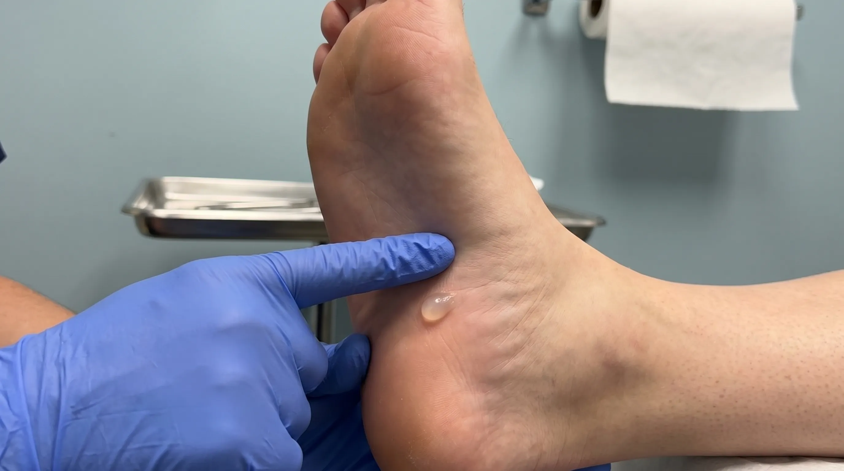
Perhaps the most crucial piece of medical information to understand is that a cyst foot bottom is entirely noncancerous. The term "tumor" is often used loosely in everyday conversation to describe any abnormal lump, but in medical terminology, a tumor simply refers to a mass or swelling. Ganglion cysts are classified as benign soft tissue lesions. They do not contain malignant cells, they do not invade neighboring structures aggressively, and they do not spread to distant parts of the body. Extensive epidemiological studies and decades of clinical observation have never linked these cysts to cancer development. The primary concern with a plantar cyst revolves around mechanical discomfort and functional limitation rather than oncological risk. Reassuring patients of this benign nature is a standard first step in clinical management, allowing individuals to focus their energy on symptom relief and lifestyle adjustments rather than catastrophic health scenarios.
Epidemiology and Primary Risk Factors
While anyone can develop a plantar cyst, certain demographic and lifestyle factors significantly increase the probability of occurrence. Understanding who is most susceptible and why helps individuals take targeted preventative measures and seek appropriate medical guidance when necessary.
Demographics: Who Is Most Susceptible
Clinical data consistently reveals a pronounced gender disparity in ganglion cyst prevalence. Women are approximately three times more likely to develop these fluid-filled sacs than men. Researchers believe this difference may be partially attributed to anatomical variations in foot structure, differences in joint laxity, hormonal influences on connective tissue elasticity, and footwear choices that place unique mechanical stresses on the forefoot and plantar fascia. Age is another critical variable. These cysts rarely appear in children and are exceptionally uncommon in elderly populations. The peak incidence occurs between the ages of twenty and fifty, aligning with the most physically active decades of human life. During this period, individuals typically engage in higher-impact recreational activities, professional labor that demands prolonged standing or walking, and athletic pursuits that place repetitive strain on the ankle and foot joints.
Occupational and Athletic Contributors
The repetitive application of mechanical stress is a well-documented catalyst for synovial tissue herniation. Professionals whose daily routines involve constant weight-bearing, sudden directional changes, or prolonged use of rigid footwear demonstrate a markedly higher incidence of a cyst foot bottom. Dancers, gymnasts, construction workers, nurses, and runners frequently experience the exact biomechanical conditions that weaken the tendon sheath and joint capsule. Gymnasts, for instance, endure extreme ankle dorsiflexion and plantarflexion, creating substantial pressure differentials within the joint space. When these forces are applied consistently over months or years, the structural integrity of the synovial lining gradually deteriorates, allowing fluid to escape and accumulate. Even seemingly minor habits, such as wearing high heels with inadequate arch support or engaging in weekend sports without proper conditioning, can contribute to the cumulative microtrauma that ultimately triggers cyst development.
The Role of Prior Injuries and Arthritis
A history of foot trauma significantly elevates long-term risk. Sprained ankles, tendon ruptures, stress fractures, and severe contusions can permanently alter the architecture of the joint capsule and surrounding connective tissue. Even after the initial injury has healed, the scarred tissue remains structurally weaker and more prone to fluid leakage under normal physiological stress. Furthermore, individuals with early-stage osteoarthritis are at notably higher risk. Degenerative joint disease causes the cartilage to wear down, increasing friction and altering the normal distribution of synovial fluid. The body attempts to compensate by producing more lubricating fluid, which can overwhelm the joint capsule and force its way outward through weakened areas. This explains why older adults with pre-existing joint degeneration occasionally notice new masses, even if they are outside the typical age range for peak incidence.
Recognizing Symptoms and Clinical Presentation
Not all plantar masses announce themselves with identical symptoms. The clinical presentation of a cyst foot bottom varies widely depending on its exact location, size, and proximity to nearby neurological structures. Learning to distinguish characteristic features from warning signs ensures timely and appropriate medical evaluation.
Visual and Tactile Characteristics
The primary and most obvious symptom is the presence of a distinct bump beneath the skin of the sole. Visually, the mass typically appears round or oval, with clearly defined borders. When palpated, it usually feels firm, similar to a small rubber ball, though some cysts may feel surprisingly soft or spongy depending on their internal pressure and the thickness of the overlying skin. One of the most defining clinical features is the mass's mobility. Unlike deeply embedded tumors or fixed bony prominences, a ganglion cyst typically shifts slightly when gentle pressure is applied, sliding within the subcutaneous tissue layer. In certain lighting conditions or when the skin is thin, the cyst may exhibit a faint translucent quality, hinting at the clear fluid trapped inside. Many patients report that the bump is initially painless, only seeking medical attention when it grows large enough to interfere with footwear or daily activities.
Size Fluctuations and Activity Correlation
One of the most fascinating aspects of a cyst foot bottom is its dynamic nature. The size of the lump rarely remains static. Patients frequently observe that the cyst enlarges during periods of intense physical activity or prolonged standing, and gradually shrinks after extended rest. This phenomenon occurs directly due to changes in joint pressure. Movement stimulates increased synovial fluid production and pushes existing fluid into the cystic sac, causing it to expand. Conversely, immobilization allows the fluid to slowly reabsorb or leak back into the joint space, reducing the visible bulge. These fluctuations can occur over days or weeks and sometimes lead patients to believe the problem is resolving itself, only for the cyst to return when normal activity resumes. Tracking these changes using a simple journal or periodic photographs provides valuable information during clinical consultations.
Nerve Compression and Neurological Symptoms
The plantar surface of the foot houses a dense network of sensory and motor nerves responsible for balance, proprioception, and tactile feedback. When a cyst expands directly over a nerve bundle or presses into the surrounding neurovascular pathway, symptoms shift dramatically. Pain becomes the dominant complaint, often described as a sharp, burning, or aching sensation that radiates along the sole. Tingling, pins-and-needles paresthesia, or localized numbness frequently accompany the pain, indicating direct neural irritation. In advanced cases where motor nerves are compromised, patients may experience subtle muscle weakness, affecting their ability to curl their toes or maintain a stable gait. Any cyst foot bottom accompanied by neurological symptoms warrants prompt medical evaluation, as prolonged nerve compression can lead to chronic discomfort and altered walking mechanics.
Occult Ganglions: The Hidden Pain Source
A particularly challenging clinical variation involves occult ganglions. These cysts are too small to produce a visible or palpable bump beneath the skin, yet they cause significant, localized pain. Because they lack a noticeable mass, patients and even primary care providers may initially struggle to identify the root cause of foot discomfort. The pain from occult ganglions often mimics plantar fasciitis, stress fractures, or neuromas. Specialized imaging becomes essential in these scenarios, as the physical examination alone cannot confirm the diagnosis. Recognizing this variant prevents unnecessary delays in treatment and spares patients from pursuing ineffective interventions for misdiagnosed conditions.
Professional Diagnosis and Differential Evaluation
Accurate identification is the cornerstone of effective management. While a cyst foot bottom is usually benign, several other conditions present with nearly identical symptoms. A systematic diagnostic approach ensures that patients receive appropriate care and that more serious pathologies are definitively ruled out.
Physical Examination and Transillumination
The diagnostic process typically begins with a thorough clinical evaluation. A podiatrist or orthopedic specialist will assess the exact location, size, consistency, and mobility of the mass while asking detailed questions about symptom onset and activity correlation. One of the most straightforward and reliable clinical tests is transillumination. Using a bright, focused penlight, the clinician shines the light directly against the cyst. If the structure contains clear synovial fluid, the light passes through easily, causing the mass to glow with a faint, translucent halo. Solid masses, such as fibromas or lipomas, block the light entirely and remain opaque. While not infallible, transillumination provides rapid, preliminary confirmation that guides subsequent diagnostic steps.
Advanced Imaging Modalities
When the clinical picture is unclear, or when surgical planning is necessary, advanced imaging becomes indispensable. Standard X-rays are routinely ordered first, even though cysts themselves do not appear on radiographic film. The primary purpose of an X-ray is to visualize underlying bone structure, rule out arthritic degeneration, stress fractures, or rare bone tumors, and assess joint alignment. If soft tissue detail is required, ultrasound or magnetic resonance imaging (MRI) is utilized. Ultrasound offers a dynamic, real-time view of the cyst, confirming its fluid-filled nature, visualizing the stalk connecting to the joint, and guiding precise needle aspiration if chosen. MRI provides the most comprehensive three-dimensional mapping, clearly differentiating a cyst foot bottom from ligamentous tears, tendon pathology, and other soft tissue neoplasms. This level of detail is particularly crucial for occult ganglions or complex cases involving nerve entrapment.
Distinguishing from Plantar Fibromas and Other Masses
Differential diagnosis is critical because management strategies vary drastically depending on the tissue composition. A plantar fibroma, for example, develops within the dense connective tissue of the plantar fascia. It feels rock-hard, does not change size with activity, and lacks the translucent quality of a ganglion cyst. Epidermoid cysts originate from trapped skin cells and often feature a central punctum, occasionally draining keratinous material. Lipomas are composed of mature fat cells, feeling remarkably soft and doughy to the touch. Bone spurs and ganglion cysts can occasionally present in similar locations, though spurs are immovable and clearly visible on X-ray. The Cleveland Clinic emphasizes that professional evaluation prevents misdiagnosis and ensures patients pursue the correct treatment pathway.
Comprehensive Treatment Pathways
Management of a cyst foot bottom follows a stepwise approach, beginning with conservative measures and progressing to procedural interventions only when symptoms significantly impair quality of life or daily function. The decision to treat is never based solely on the presence of the lump, but rather on its impact on mobility and comfort.
Non-Surgical Management Strategies
The majority of clinical guidelines, including those from the American Academy of Orthopaedic Surgeons (AAOS), strongly recommend non-invasive management as the first line of defense. Since many cysts cause minimal discomfort and occasionally resolve spontaneously, aggressive intervention is often unnecessary.
Watchful Waiting and Symptom Monitoring
When a plantar cyst is painless and does not interfere with footwear or gait, observation is a medically sound and widely accepted approach. Patients are educated on normal cyst behavior, taught to monitor size fluctuations, and advised to avoid activities that exacerbate pressure. Regular self-examinations help track progression without medical intervention. Studies indicate that a significant percentage of ganglion cysts diminish in size or disappear entirely over several months to years as the underlying tissue tear heals and the stalk naturally obliterates.
Immobilization and Activity Modification
For cysts that cause intermittent discomfort, temporary immobilization can be highly effective. A rigid brace, walking boot, or specialized orthotic splint restricts excessive joint movement and reduces the mechanical pumping action that pushes fluid into the cyst. By stabilizing the affected joint, synovial pressure normalizes, allowing the fluid to gradually reabsorb. Activity modification plays a complementary role. Patients are encouraged to switch to low-impact exercises such as swimming or stationary cycling, avoid prolonged standing on hard surfaces, and take scheduled rest periods during demanding work shifts. This conservative strategy successfully reduces cyst volume in a substantial number of patients without surgical risk.
Aspiration Procedure: Benefits and Limitations
When conservative measures fail to provide adequate relief, clinicians may recommend needle aspiration. Performed in a clinical setting under local anesthesia, a sterile needle is inserted directly into the cystic sac. The practitioner carefully withdraws the thick, gelatinous fluid, often injecting a mild corticosteroid to reduce inflammation and inhibit further fluid production. While aspiration offers rapid decompression and immediate symptom relief, it has notable limitations. The procedure does not remove the stalk connecting the cyst to the joint, meaning the underlying structural defect remains. Clinical data reveals a recurrence rate ranging from fifty to seventy percent following aspiration. Consequently, many specialists reserve this intervention for patients who prefer a quick, minimally invasive option or who are poor surgical candidates, fully informing them of the high likelihood of return.
Surgical Intervention: Ganglionectomy
For persistent, painful, or recurrent cysts that significantly compromise daily function, surgical excision represents the definitive solution.
Preoperative Considerations
Before proceeding with surgery, a thorough risk-benefit discussion is essential. Patients are informed about the procedure, expected recovery timeline, potential complications, and recurrence rates. Preoperative imaging ensures precise localization of the cyst and its stalk. Individuals with underlying conditions such as diabetes, peripheral vascular disease, or compromised immune function require specialized preoperative optimization to minimize infection risk and promote optimal wound healing.
The Procedure Explained
Ganglionectomy is typically performed as an outpatient procedure under regional or general anesthesia. The surgeon makes a small, strategically placed incision on the sole of the foot to access the cyst while avoiding critical neurovascular structures. Once exposed, the cyst is carefully dissected free from surrounding tissues. The critical step involves tracing the narrow stalk back to its origin at the joint capsule or tendon sheath and meticulously ligating or excising the connection to prevent fluid reaccumulation. The wound is closed in layers using absorbable sutures, and a protective dressing or boot is applied immediately.
Postoperative Recovery and Rehabilitation
Recovery follows a structured timeline. The first forty-eight to seventy-two hours focus on elevation and ice to control swelling and manage pain. Patients use crutches or a surgical shoe to limit weight-bearing pressure on the incision site. Suture removal occurs approximately two to three weeks post-procedure. As the wound heals, gentle range-of-motion exercises and gradual weight-bearing progressions are introduced. Physical therapy plays a vital role in restoring proprioception, strengthening intrinsic foot muscles, and ensuring a smooth return to normal footwear. Most patients achieve full functional recovery within six to eight weeks, with long-term success rates exceeding eighty percent.
Daily Management, Footwear, and Self-Care
Living with a cyst foot bottom requires thoughtful adjustments to daily routines. Implementing evidence-based self-care strategies minimizes discomfort, prevents exacerbation, and supports overall foot biomechanics.
Choosing Supportive Footwear and Orthotics
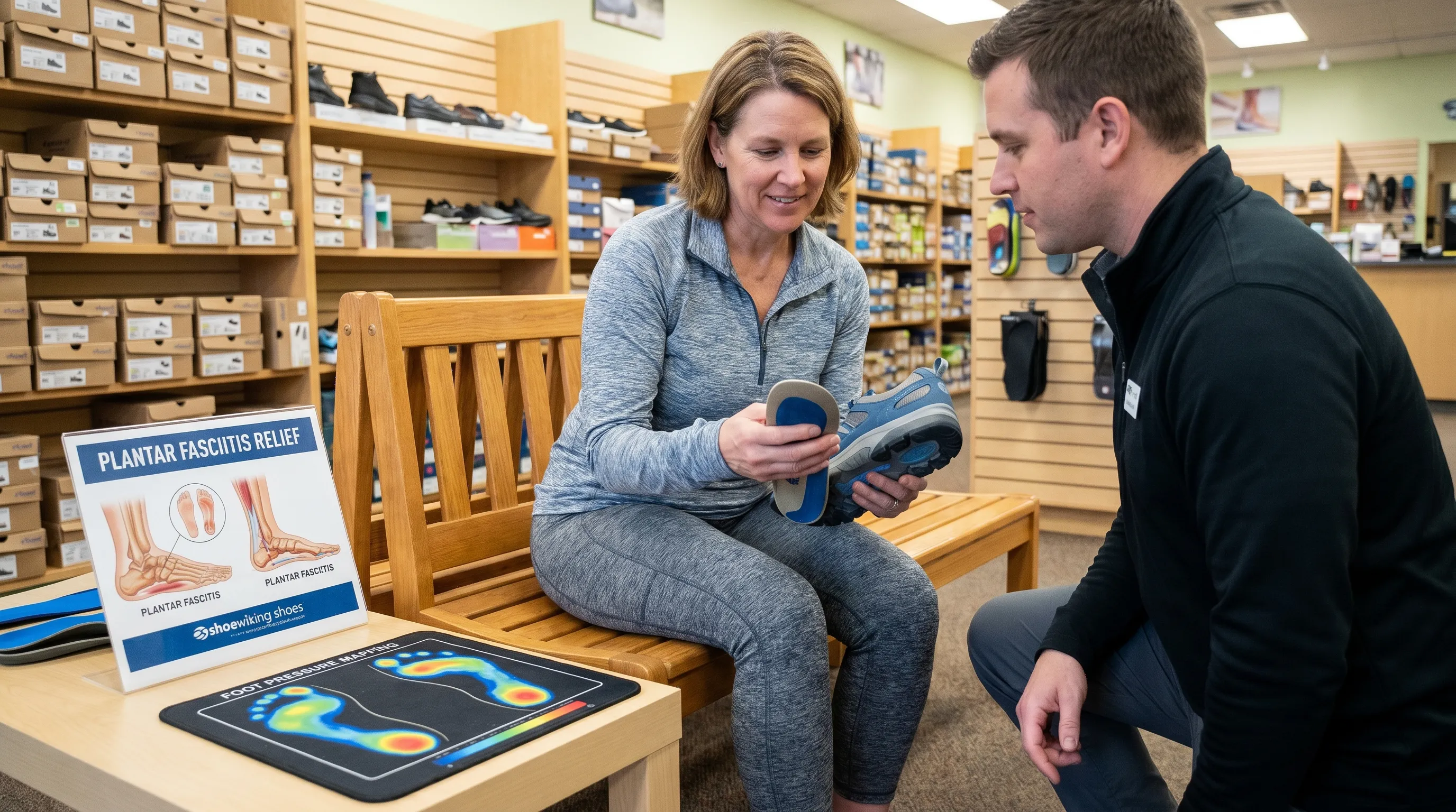
Footwear selection is arguably the most impactful lifestyle factor. Shoes with narrow toe boxes, thin soles, or inadequate arch support concentrate pressure directly onto the plantar surface, irritating the cyst and stimulating further fluid production. Opting for shoes with a wide forefoot, cushioned midsoles, and removable insoles allows for custom orthotic placement. Over-the-counter or prescription orthotics can redistribute weight away from the cystic area, creating a protective pocket that eliminates direct friction. The Mayo Clinic consistently highlights the importance of proper footwear in managing plantar masses.
Pressure Relief Techniques for Walking
For individuals required to stand or walk extensively for work, strategic pressure relief techniques can dramatically improve comfort. Using gel pads, foam cushions, or custom-cut felt donuts placed adjacent to (not directly over) the cyst helps disperse impact forces. Rotating between two pairs of supportive shoes throughout the week prevents repetitive stress on identical pressure points. Implementing micro-breaks every hour to remove shoes, elevate the feet, and gently stretch the plantar fascia reduces intra-articular pressure and limits cyst expansion. These simple, cost-effective modifications are highly effective adjuncts to medical treatment.
Home Monitoring and Red Flags
While self-management is encouraged, patients must remain vigilant for warning signs that necessitate immediate professional evaluation. Sudden, severe pain; rapid, unexplained enlargement; skin changes such as redness, warmth, or drainage; and progressive numbness or tingling extending into the toes indicate potential complications like infection, rupture, or advanced nerve compression. Documenting symptoms with a simple log helps clinicians track progression and adjust treatment plans efficiently. Avoiding folk remedies such as smashing the cyst with heavy objects is critical, as this causes severe tissue trauma, inflammation, and significantly increases infection risk without eliminating the underlying stalk.
| Feature | Ganglion Cyst (Cyst Foot Bottom) | Plantar Fibroma | Epidermoid Cyst | Lipoma |
|---|---|---|---|---|
| Tissue Composition | Synovial fluid in fibrous sac | Dense collagenous scar tissue | Trapped skin cells and keratin | Mature adipose (fat) tissue |
| Consistency on Palpation | Firm to spongy, slightly mobile | Rock-hard, fixed to fascia | Firm, often with central punctum | Soft, doughy, highly mobile |
| Size Fluctuation | Changes with activity/rest | Remains constant | Grows slowly over years | Grows slowly over years |
| Transillumination | Glows with light penetration | Opaque, blocks light | Opaque | Opaque or partially translucent |
| Primary Management | Observation, aspiration, excision | Orthotics, injections, rarely surgery | Surgical excision if symptomatic | Observation or elective excision |
Frequently Asked Questions
What exactly causes a cyst foot bottom to develop?
A cyst foot bottom typically develops due to a small tear in the tissue surrounding a joint or tendon sheath. This allows thick, joint-lubricating synovial fluid to leak out and pool, forming a sac that bulges beneath the skin. Factors like repetitive stress, prior joint injuries, and underlying osteoarthritis significantly increase the likelihood of formation.
Is a cyst on the bottom of the foot cancerous?
No. Ganglion cysts, which are the most common type found on the plantar surface, are entirely benign and noncancerous. They do not metastasize or invade surrounding tissues. However, any new or growing mass should be evaluated by a healthcare provider to rule out other soft tissue conditions.
Can I safely drain a cyst foot bottom at home?
Absolutely not. Attempting to drain a cyst at home with needles or sharp objects carries a severe risk of infection, nerve damage, and incomplete fluid removal. Professional aspiration must be performed under sterile conditions, and even then, recurrence is common without addressing the underlying stalk or joint connection.
How long does it take to recover after cyst removal surgery on the foot?
Initial wound healing typically takes two to three weeks. However, full recovery and a return to normal weight-bearing activities usually require four to eight weeks. Patients often use protective footwear, compression, and physical therapy during this period to restore mobility and prevent recurrence.
Will changing my shoes make a cyst foot bottom go away?
While footwear modifications do not eliminate the cyst itself, properly fitted shoes with ample toe box space, arch support, and cushioned soles significantly reduce pressure on the plantar surface. This can shrink cyst size, alleviate pain, and improve daily function while you monitor or pursue definitive treatment.
What is the difference between a ganglion cyst and a plantar fibroma?
A ganglion cyst is a fluid-filled sac that often changes size and may feel slightly compressible, whereas a plantar fibroma is a firm, solid nodule of scar-like tissue that develops within the plantar fascia. Fibromas do not shrink with rest, lack the translucent quality of cysts, and require different management approaches.
Conclusion
Discovering a lump on the sole can be unsettling, but understanding the nature of a cyst foot bottom reveals a highly manageable, benign condition rooted in normal physiological processes rather than severe disease. Ganglion cysts on the plantar surface arise from minor connective tissue tears, filling with joint fluid and occasionally causing discomfort when they press on nerves or interfere with weight-bearing. Through careful clinical evaluation, precise differential diagnosis, and a tiered treatment approach ranging from watchful waiting to specialized surgical intervention, the vast majority of individuals achieve complete symptom relief and restored mobility. Prioritizing proper footwear, implementing targeted activity modifications, and partnering with a qualified podiatrist or orthopedic specialist ensures optimal outcomes. By dismissing harmful myths, avoiding unsafe home remedies, and relying on evidence-based medical guidance, patients can confidently navigate their recovery and return to active, pain-free movement. For personalized advice tailored to your specific biomechanics and lifestyle, always consult a licensed healthcare professional who can provide accurate imaging, definitive diagnosis, and a customized treatment roadmap.
For additional educational resources on foot anatomy and soft tissue management, visit the American Society for Surgery of the Hand or review the comprehensive guidelines provided by the Cleveland Clinic and Mayo Clinic. Educational video content on foot cyst diagnosis and podiatric treatment approaches can also be found by searching reputable medical channels for clinical demonstrations and patient education materials.
How we work
Researched and drafted with AI assistance, then reviewed by a human editor against the cited sources.
This article is general health information, not medical advice. Consult a qualified healthcare professional about your own situation.