Pain After Root Canal: Recovery Timeline, Management Strategies, and Warning Signs
Undergoing endodontic therapy is one of the most effective ways to preserve a severely damaged tooth, yet many patients approach the procedure with understandable apprehension about post-operative discomfort. The modern reality of endodontics, as detailed by the National Institute of Dental and Craniofacial Research (NIDCR), has dramatically improved over the decades, with advanced instrumentation, localized anesthesia protocols, and biocompatible sealing materials making the treatment itself highly tolerable. However, the biological healing process that follows inevitably triggers a physiological response in the surrounding tissues. Understanding pain after root canal therapy is essential for setting realistic expectations, managing symptoms effectively, and recognizing when clinical follow-up is necessary. This comprehensive guide breaks down the science of endodontic recovery, evidence-based pain management strategies, dietary modifications, and the critical warning signs that distinguish normal tissue repair from potential complications. By following clinically validated protocols, you can navigate the healing window with confidence and return to full oral function as quickly as possible.
Understanding Post-Root Canal Discomfort: Why It Happens
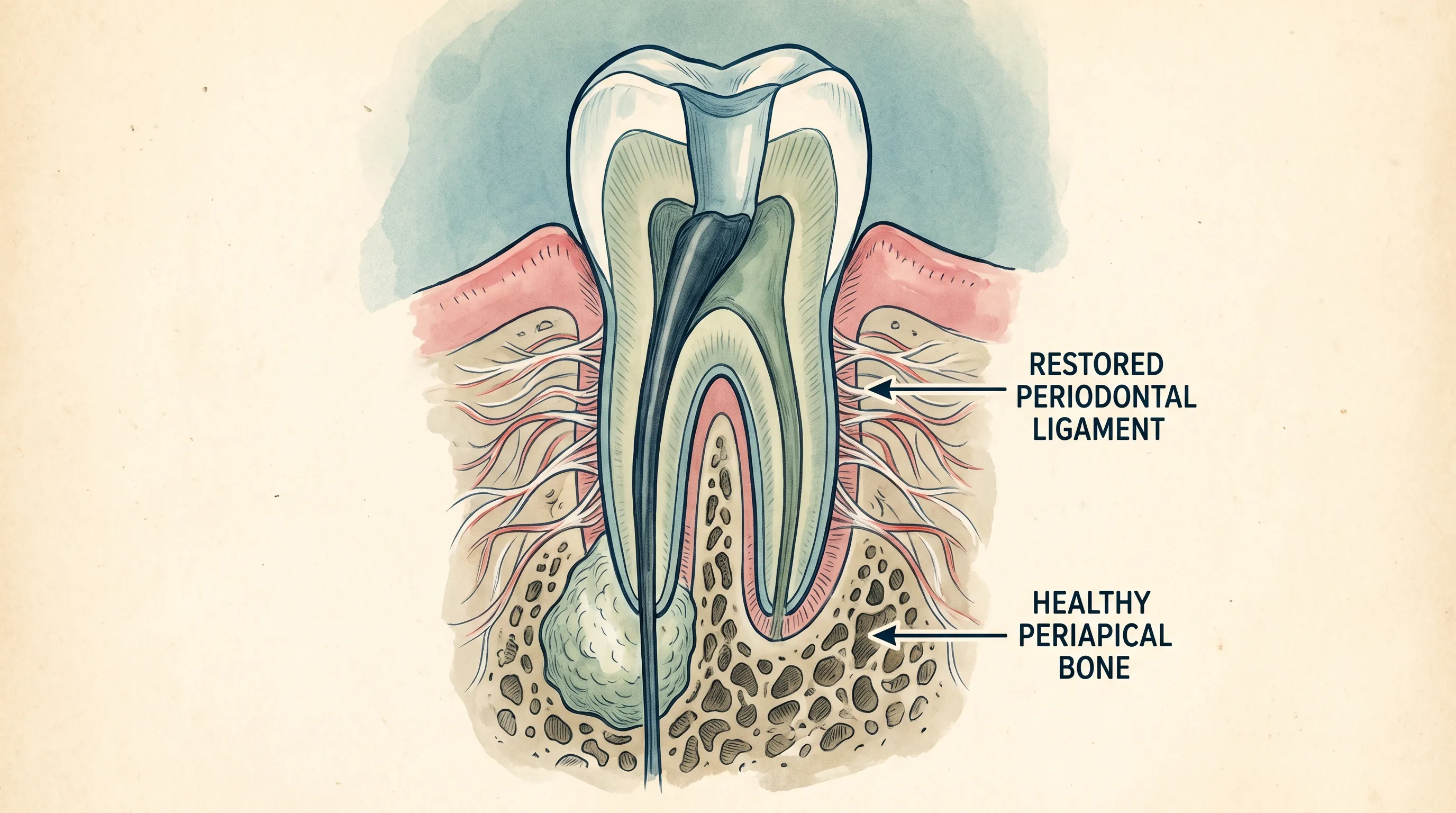
Tooth preservation procedures fundamentally alter the internal anatomy of a dental structure, and the body responds predictably to this controlled intervention. During endodontic therapy, the inflamed or necrotic pulp tissue is removed, the root canal system is meticulously cleaned, shaped, and disinfected, and then sealed with gutta-percha and bioceramic sealers. Even with flawless technique, the mechanical instrumentation and chemical irrigation necessary to eliminate pathogens cause localized tissue trauma. This trauma initiates a cascade of inflammatory mediators, including prostaglandins, bradykinin, and interleukins, which sensitize nociceptors in the periodontal ligament and periapical bone. The resulting discomfort is not a sign of procedural failure but rather a natural component of the body's reparative response.
Free calculatorHeadache Location ChartIdentify headache types by locationOpen the calculatorThe Biological Mechanism Behind Healing
The periodontal ligament acts as a microscopic shock absorber that anchors the tooth root to the alveolar bone. This ligament is densely populated with nerve fibers and vascular networks. When endodontic files extend slightly beyond the apical foramen during cleaning, or when irrigating solutions like sodium hypochlorite interact with the apical tissues, transient inflammation occurs. This inflammation increases interstitial fluid pressure, compressing nerve endings and creating the sensation of tenderness upon biting or percussion. Additionally, the immune system mobilizes macrophages and fibroblasts to clear residual debris and initiate collagen deposition. This cellular activity peaks within forty-eight to seventy-two hours, aligning precisely with the most intense phase of post-operative sensitivity. Recognizing that pain after root canal treatment is fundamentally driven by acute inflammatory resolution rather than persistent infection helps patients approach recovery with reduced anxiety and better compliance with aftercare instructions.
Inflammation and Nerve Irritation Explained
Neurogenic inflammation plays a critical role in endodontic discomfort. The dental pulp, once removed, is no longer the source of pain; however, the periradicular tissues take center stage during healing. Sympathetic nerve fibers in the periapical region release neuropeptides such as substance P and calcitonin gene-related peptide (CGRP), which dilate blood vessels and increase vascular permeability. This leads to localized edema and heightened sensitivity to thermal stimuli, mechanical pressure, and occlusal forces. The brain interprets these signals as aching or throbbing discomfort, often radiating to adjacent teeth, the jawline, or even the ear due to the shared trigeminal nerve pathways. This referred sensation is entirely normal and typically diminishes as prostaglandin synthesis is regulated and tissue homeostasis is restored. Patients should be aware that nerve irritation can occasionally mimic a headache or sinus pressure, particularly in maxillary molars where roots sit in close proximity to the maxillary sinus floor.
Typical Recovery Timeline: What to Expect Day by Day
Endodontic healing follows a predictable trajectory when the procedure is performed successfully and post-operative care is strictly followed. As outlined by the Cleveland Clinic, while individual variability exists based on factors like baseline inflammation severity, systemic health conditions, and anatomical complexity, most patients follow a standardized recovery curve. Monitoring your symptoms against this timeline provides valuable insight into whether your body is responding appropriately or if clinical intervention is warranted.
First 24–48 Hours: The Acute Phase
The initial twenty-four to forty-eight hours represent the peak of inflammatory activity. Patients commonly report moderate aching, sensitivity to percussion, and a feeling of heaviness or fullness around the treated tooth. Mild gingival tenderness is also typical, especially if a rubber dam clamp was positioned deeply during the procedure. During this window, the local anesthetic has completely dissipated, and vasoconstriction has reversed, leading to maximal blood flow and cellular recruitment to the surgical site. Swelling is usually minimal but can occasionally present as mild facial puffiness, particularly in cases with pre-existing periapical pathology. Strict adherence to prescribed analgesics, ice application protocols, and dietary modifications significantly mitigates acute discomfort. Avoidance of hot beverages and hard chewing is non-negotiable during this phase to prevent exacerbating ligament inflammation.
Days 3–7: Gradual Improvement
By the third day, the acute inflammatory cascade transitions into the proliferative healing stage. Prostaglandin levels drop, macrophage activity decreases, and fibroblasts begin organizing new connective tissue fibers within the periodontal ligament. Pain after root canal treatment typically shifts from constant throbbing to intermittent tenderness that occurs primarily with biting pressure or aggressive brushing. Most patients can safely transition to a semi-soft diet and gradually resume normal oral hygiene routines, though vigorous rinsing or electric toothbrush use should still be moderated. Any residual numbness or tingling beyond this timeframe should be documented and reported, as it may indicate transient nerve irritation or anatomical variation. The psychological relief that often accompanies this improvement phase reinforces the effectiveness of endodontic therapy when managed correctly.
Weeks 2–4: Complete Healing Phase
During the second to fourth week, periapical bone remodeling accelerates. Osteoclasts resorb micro-fractures caused by instrumentation, while osteoblasts deposit new trabecular bone to stabilize the apical seal. At this stage, the tooth should feel indistinguishable from adjacent healthy teeth during normal function. Occasional mild sensitivity to extreme temperature changes or heavy biting forces may persist in heavily calcified canals or multi-rooted teeth with complex anatomy. Follow-up radiographic evaluation is often scheduled during this window to confirm apical resolution and rule out delayed flare-ups. Patients who have adhered to their recovery protocol typically experience zero limitations in chewing, speaking, or maintaining hygiene. The final permanent restoration, usually a porcelain-fused-to-metal or full-zirconia crown, is placed to protect the structurally compromised tooth from fracture and restore full occlusal harmony.
Distinguishing Normal Pain After Root Canal From Warning Signs
While discomfort is expected, not all post-endodontic sensations indicate healthy tissue repair. Clinical research emphasizes that early identification of abnormal pain significantly improves outcomes and prevents unnecessary tooth loss. Patients must learn to differentiate between routine inflammatory soreness and symptoms that suggest procedural complications, anatomical challenges, or secondary infection. The Mayo Clinic provides clear clinical benchmarks for evaluating recovery progress and identifying potential dental infections.
When Discomfort is Part of the Healing Process
Normal pain after root canal therapy remains localized, gradually diminishes in intensity, and responds predictably to over-the-counter analgesics. Tenderness when tapping the tooth lightly or biting on a cotton roll is expected for five to seven days. Mild gum soreness near the rubber dam attachment sites, slight jaw stiffness from keeping the mouth open during the procedure, and occasional throbbing when lying flat are all within standard parameters. The pain should never wake you from sleep, radiate uncontrollably down the neck, or produce visible pus drainage. If your symptoms follow a steady downward trajectory and you can maintain hydration, sleep, and light activity, your body is likely executing a textbook inflammatory resolution. Consistency in medication timing and avoiding occlusal trauma on the treated side are the most effective ways to support this natural progression.
Red Flags That Require Immediate Professional Attention
Certain symptoms demand prompt clinical evaluation and potentially adjunctive intervention. Persistent or escalating pain beyond ten days, spontaneous throbbing that disrupts sleep, or sharp pain upon releasing biting pressure are hallmark indicators of a high occlusal restoration or vertical root fracture. Facial swelling that expands toward the eye, neck, or floor of the mouth suggests a spreading cellulitis that requires antibiotic therapy and possible drainage, a scenario closely monitored by the CDC for dental infection complications. Fever above 101°F, foul-tasting drainage from the gingival margin, or a mobile tooth structure indicates re-infection or loss of the coronal seal. Additionally, prolonged numbness in the lower lip or chin following mandibular procedures may indicate inferior alveolar nerve proximity or inflammation requiring corticosteroid management. Ignoring these red flags risks irreversible damage, systemic infection, or the need for tooth extraction. Early communication with your endodontic provider ensures accurate diagnosis through percussion testing, radiographic evaluation, and occlusal adjustment.
Evidence-Based Strategies for Managing Pain After Root Canal
Effective symptom management combines pharmacological intervention, targeted physical therapies, and behavioral modifications. Decades of clinical trials have established clear protocols for minimizing endodontic discomfort while supporting optimal tissue regeneration. Patients who proactively implement these strategies experience shorter recovery periods and higher satisfaction rates.
Over-the-Counter Medications and Dosing Guidelines
The cornerstone of post-endodontic analgesia is a staggered regimen of non-steroidal anti-inflammatory drugs (NSAIDs) combined with acetaminophen. Ibuprofen and acetaminophen guidelines from the NIH note that proper dosing significantly inhibits cyclooxygenase enzymes, reducing prostaglandin synthesis and periapical edema. Ibuprofen dosed at 400 to 600 milligrams every six hours works synergistically with acetaminophen at 500 to 650 milligrams every four to six hours, which complements this by acting centrally on pain pathways. Clinical studies demonstrate that alternating these medications every three hours provides superior relief compared to either agent alone, without exceeding hepatic or gastrointestinal safety thresholds. Patients with NSAID allergies, peptic ulcers, or renal impairment should rely on acetaminophen monotherapy and consult their prescribing physician. Opioids are rarely indicated for routine endodontic recovery and are reserved for severe, acute exacerbations unresponsive to first-line treatments. Always take analgesics with food, maintain strict adherence to maximum daily limits, and discontinue use once discomfort subsides to a manageable baseline.
Natural and Home-Based Pain Relief Techniques
Pharmacological management should be paired with conservative home therapies that accelerate tissue recovery and reduce muscular tension. Applying an external ice pack to the cheek over the treated area for fifteen minutes on, followed by fifteen minutes off, effectively constricts superficial blood vessels and limits inflammatory mediators from pooling. After forty-eight hours, transitioning to warm compresses promotes vasodilation and lymphatic drainage. Elevated sleeping positions using two pillows prevent nocturnal venous congestion that exacerbates throbbing sensations. Gentle saltwater rinses (one-half teaspoon of salt dissolved in eight ounces of warm water) maintain microbial balance, soothe gingival irritation, and prevent food debris accumulation near the temporary filling. Stress reduction through deep diaphragmatic breathing and progressive muscle relaxation also lowers cortisol levels, which has been shown to improve immune efficiency and pain tolerance during the critical first week of healing.
Common Complications and Why Pain May Persist
While modern endodontics boasts success rates exceeding ninety percent, anatomical complexities and biological variables can occasionally lead to prolonged symptoms. Understanding these potential complications empowers patients to engage in informed discussions with their dental providers and seek timely diagnostics when recovery deviates from expected norms.
Missed Canals and Incomplete Cleaning
The root canal system is far more intricate than traditional two-dimensional radiographs suggest. Maxillary molars frequently contain a hidden fourth canal, the MB2 (mesiobuccal second), located beneath the primary mesiobuccal canal. If missed during instrumentation, residual necrotic tissue and bacterial biofilms remain, perpetuating apical inflammation and chronic discomfort. Mandibular incisors occasionally feature dual canals, and premolars may exhibit C-shaped anatomies in Asian populations that defy standard cleaning protocols. Cone-beam computed tomography (CBCT) has become the gold standard for detecting these variations before symptoms escalate. When pain after root canal therapy persists despite proper analgesic use, endodontic retreatment utilizing ultrasonic activation, surgical microscopy, and bioceramic obturation typically resolves the issue by addressing the untouched anatomical territory.
Cracked Tooth Structure and Fractures
Teeth requiring endodontic intervention have often endured years of heavy occlusal loading, large restorations, and bruxism, leaving them structurally compromised. Hairline fractures that extend vertically through the root or horizontally across the cervical third can develop during instrumentation or shortly after temporary crown placement. These cracks allow bacterial infiltration and prevent the periodontal ligament from healing, resulting in localized pain upon chewing or temperature exposure. Diagnosis often requires transillumination, bite stick testing, and careful radiographic evaluation of the periodontal space. Once a vertical root fracture is confirmed, the prognosis shifts dramatically, as the fractured segment cannot be sealed biologically. Extraction and implant replacement become the standard of care to prevent progressive bone loss and chronic infection. Early detection through functional limitations and targeted percussion sensitivity is crucial for preserving remaining dentition.
Re-Infection and Delayed Crown Placement
The temporary filling material placed after endodontic cleaning serves as a microbial barrier, but it degrades over time and does not withstand occlusal forces. Prolonged delay in permanent crown placement allows salivary bacteria to penetrate micro-gaps, recontaminating the sealed canals and triggering secondary periapical abscess formation. Additionally, coronal leakage compromises the hermetic seal achieved with gutta-percha and resin sealer, allowing endotoxins like lipopolysaccharides to leach into the surrounding bone. Patients who postpone final restoration beyond three weeks face significantly higher risks of flare-ups, necessitating root canal retreatment or apicoectomy surgery. Ensuring prompt fabrication and cementation of a properly contoured crown not only eliminates microbial pathways but also distributes biting forces evenly, protecting the fragile tooth structure from catastrophic fracture.
Dietary and Lifestyle Adjustments for Faster Recovery
Nutritional support and behavioral modifications play an underappreciated role in endodontic healing. The foods you consume, the substances you avoid, and your daily habits directly influence inflammatory resolution, tissue regeneration, and immune function. Implementing strategic adjustments during the recovery window minimizes mechanical stress and optimizes the biological environment for rapid periapical repair.
Foods to Embrace and Avoid
A modified soft diet should be maintained for at least five to seven days post-procedure. Opt for nutrient-dense, easily masticable options such as oatmeal, mashed sweet potatoes, scrambled eggs, Greek yogurt, lukewarm bone broths, and blended smoothies fortified with protein powder and omega-3 fatty acids. These foods provide essential amino acids, zinc, vitamin C, and anti-inflammatory compounds that accelerate collagen synthesis and bone remodeling. Strictly avoid hard, crunchy, or sticky foods like nuts, raw carrots, ice, caramel, and tough meats that require forceful chewing. Extremely hot or cold items should be minimized, as thermal extremes can trigger fluid movement within microscopic dentinal tubules and provoke sharp, transient sensitivity. Chew exclusively on the contralateral side, swallow without prolonged mastication, and maintain hydration to support salivary buffering capacity and tissue perfusion.
Oral Hygiene Practices During the Healing Window
Maintaining meticulous oral hygiene prevents secondary plaque accumulation and gingival inflammation that can mask or exacerbate endodontic discomfort. Continue brushing twice daily with a soft-bristled toothbrush, but use gentle, circular motions around the treated tooth to avoid disrupting the temporary restoration or irritating healing gingival margins. Flossing should be performed carefully, pulling the thread straight out rather than snapping it back to avoid dislodging the provisional filling. Antimicrobial mouthwashes containing cetylpyridinium chloride or essential oil blends can be used sparingly if recommended by your provider, though alcohol-based rinses should be avoided due to their desiccating effects on mucosal tissue. Do not use water flossers or high-pressure oral irrigators on the treated arch until permanent restoration is complete, as directed spray pressure can force bacteria into open micro-gaps. Consistency in gentle, targeted hygiene protocols ensures the surgical site remains pristine while systemic healing progresses undisturbed.
| Symptom Category | Normal Healing Indicators | Complication Warning Signs |
|---|---|---|
| Pain Intensity | Mild to moderate, steadily decreasing | Severe, escalating, or sleep-disrupting |
| Biting Sensitivity | Tenderness on pressure, resolves by day 7 | Sharp pain on release, mobility, or heaviness |
| Swelling | Minimal or localized to gum tissue | Expanding facial puffiness, neck tightness, or visible pus |
| Temperature Response | Brief sensitivity to extremes, improves daily | Persistent throbbing with heat/cold, radiating pain |
| Healing Timeline | Full resolution within 7-10 days | Discomfort persists beyond 14 days or fluctuates |
Frequently Asked Questions
How long does pain after root canal treatment typically last?
Most patients experience mild to moderate discomfort for two to three days following a root canal. By the end of the first week, sensitivity usually resolves as the periapical tissues heal. If pain persists beyond seven to ten days, it warrants professional evaluation to rule out occlusal trauma, missed anatomy, or delayed infection.
Is severe pain after a root canal considered normal?
Severe, throbbing, or escalating pain is not typical and may indicate complications such as an infection flare-up, missed canal, occlusal trauma from a high restoration, or a vertical root fracture. Contact your endodontist promptly if pain intensifies or disrupts sleep.
What is the best over-the-counter medication for post-root canal pain?
A combination of ibuprofen and acetaminophen is widely recommended by dental professionals for optimal endodontic pain relief. Ibuprofen reduces inflammatory swelling while acetaminophen targets central pain pathways. Always follow dosing guidelines and consult your dentist if you have contraindications.
Can I chew on a tooth that just had a root canal?
You should avoid chewing on the treated tooth until it has been permanently restored with a crown. The tooth structure is temporarily weakened during endodontic therapy, and premature chewing can cause fractures, microleakage, or delayed healing of the surrounding ligament.
Why does a tooth sometimes hurt months after a root canal?
Delayed pain can result from a cracked root, persistent bacterial contamination, an untreated accessory canal, a failed coronal seal, or adjacent periodontal disease. Advanced imaging and clinical percussion testing help identify the source and determine whether retreatment or extraction is necessary.
Should I use ice or heat to manage post-root canal discomfort?
Ice is recommended during the first forty-eight hours to reduce inflammation and numb sharp pain. After the initial swelling subsides, moist heat can help improve blood flow and relax tense jaw muscles. Avoid direct ice on the gums and limit cold packs to fifteen-minute intervals.
How do I know if my temporary filling has failed?
Signs of a compromised temporary filling include a rough or missing surface, increased sensitivity to temperature, food impaction around the tooth, or a persistent bad taste. If you notice these changes, schedule a prompt evaluation to prevent bacterial re-infiltration and potential canal contamination.
Can stress make pain after root canal therapy worse?
Yes, elevated cortisol and adrenaline levels from chronic stress can heighten nociceptor sensitivity and prolong inflammatory resolution. Practicing mindfulness, adequate sleep, and hydration helps modulate the nervous system's pain response and accelerates endodontic recovery.

Conclusion
Navigating the recovery window following endodontic therapy requires a blend of clinical understanding, disciplined aftercare, and timely communication with dental professionals. Pain after root canal treatment is overwhelmingly a predictable, self-limiting inflammatory response that signals healthy tissue regeneration rather than procedural failure. By adhering to evidence-based analgesic protocols, implementing strategic dietary modifications, and maintaining gentle yet consistent oral hygiene, patients can significantly accelerate periapical healing and restore full occlusal function. Recognizing the boundary between normal post-operative sensitivity and clinical red flags empowers individuals to seek appropriate intervention before minor complications escalate into irreversible damage. Modern endodontics continues to refine instrumentation precision, biocompatible sealers, and three-dimensional imaging, ensuring that tooth preservation remains a highly successful and minimally uncomfortable journey. When approached with patience, proper medication management, and realistic expectations, the path to long-term dental health is smooth, predictable, and entirely achievable.
How we work
Researched and drafted with AI assistance, then reviewed by a human editor against the cited sources.
This article is general health information, not medical advice. Consult a qualified healthcare professional about your own situation.